Tuesday, 25 March 2014

Phone Service Phone Wallpapers Icon Backgrounds Cases Wallpapers Hd Logo Call Numbers Booth Symbol Images Phones

Phone Service Biography


Source(google.com.pk)
The telegraph and telephone are both wire-based electrical systems, and Alexander Graham Bell's success with the telephone came as a direct result of his attempts to improve the telegraph.
When Bell began experimenting with electrical signals, the telegraph had been an established means of communication for some 30 years. Although a highly successful system, the telegraph, with its dot-and-dash Morse code, was basically limited to receiving and sending one message at a time. Bell's extensive knowledge of the nature of sound and his understanding of music enabled him to conjecture the possibility of transmitting multiple messages over the same wire at the same time. Although the idea of a multiple telegraph had been in existence for some time, Bell offered his own musical or harmonic approach as a possible practical solution. His "harmonic telegraph" was based on the principle that several notes could be sent simultaneously along the same wire if the notes or signals differed in pitch.


Alexander Graham Bell - Talk with Electricity

By October 1874, Bell's research had progressed to the extent that he could inform his future father-in-law, Boston attorney Gardiner Greene Hubbard, about the possibility of a multiple telegraph. Hubbard, who resented the absolute control then exerted by the Western Union Telegraph Company, instantly saw the potential for breaking such a monopoly and gave Bell the financial backing he needed. Bell proceeded with his work on the multiple telegraph, but he did not tell Hubbard that he and Thomas Watson, a young electrician whose services he had enlisted, were also exploring an idea that had occurred to him that summer - that of developing a device that would transmit speech electrically.
While Alexander Graham Bell and Thomas Watson worked on the harmonic telegraph at the insistent urging of Hubbard and other backers, Bell nonetheless met in March 1875 with Joseph Henry, the respected director of the Smithsonian Institution, who listened to Bell's ideas for a telephone and offered encouraging words. Spurred on by Henry's positive opinion, Bell and Watson continued their work. By June 1875 the goal of creating a device that would transmit speech electrically was about to be realized. They had proven that different tones would vary the strength of an electric current in a wire. To achieve success they therefore needed only to build a working transmitter with a membrane capable of varying electronic currents and a receiver that would reproduce these variations in audible frequencies.


Phone Service Phone Wallpapers Icon Backgrounds Cases Wallpapers Hd Logo Call Numbers Booth Symbol Images Phones

Phone Service Phone Wallpapers Icon Backgrounds Cases Wallpapers Hd Logo Call Numbers Booth Symbol Images Phones

Phone Service Phone Wallpapers Icon Backgrounds Cases Wallpapers Hd Logo Call Numbers Booth Symbol Images Phones

Phone Service Phone Wallpapers Icon Backgrounds Cases Wallpapers Hd Logo Call Numbers Booth Symbol Images Phones

Phone Service Phone Wallpapers Icon Backgrounds Cases Wallpapers Hd Logo Call Numbers Booth Symbol Images Phones

Phone Service Phone Wallpapers Icon Backgrounds Cases Wallpapers Hd Logo Call Numbers Booth Symbol Images Phones

Phone Service Phone Wallpapers Icon Backgrounds Cases Wallpapers Hd Logo Call Numbers Booth Symbol Images Phones

Phone Service Phone Wallpapers Icon Backgrounds Cases Wallpapers Hd Logo Call Numbers Booth Symbol Images Phones

Phone Service Phone Wallpapers Icon Backgrounds Cases Wallpapers Hd Logo Call Numbers Booth Symbol Images Phones

Phone Service Phone Wallpapers Icon Backgrounds Cases Wallpapers Hd Logo Call Numbers Booth Symbol Images Phones

Phone Service Phone Wallpapers Icon Backgrounds Cases Wallpapers Hd Logo Call Numbers Booth Symbol Images Phones

Phone Lookup Phone Wallpapers Icon Backgrounds Cases Wallpapers Hd Logo Call Numbers Booth Symbol Images Phones

Phone Lookup Biography

Source(google.com.pk)
Looking for a phone number? Here is a list of useful sites to help you.
411 Locate Reverse Phone Lookup
411 Locate provides several reverse lookup options. You may search by residential or business phone number, residential or business street address, zip code, city/state (for a zip or area code), or area code. Results include public telephone listings complete with name and address. See also 411 Locate White Pages.
411 Locate White Pages
411 Locate offers a search tool for finding "white pages" listings. Search by name to find the name, address and public telephone listing. Special features include a printer-friendly page for printing search results, an e-mail feature for sending the results to someone else, additional phone details (line type and provider), area code map and neighbor search. (See also: 411 Locate Reverse Phone Lookup)
AnyWho
This service, provided by AT&T, lets you look up public telephone numbers for businesses and residences. Search by name or business category, or use the reverse lookup feature to find the name behind a public telephone number.
Results include name and address with links to external Web sites for more search options. Results with no matches include a link to Intelius' fee-based service for more options.
Argali White & Yellow
Argali White & Yellow is software that aggregates telephone, zip code, weather and other information from sources, such as AnyWho, InfoSpace, MelissaData, Verizon Superpages and Weather Underground. The free version is supported by advertising, but the advertising does not distract from your research.
You may opt for a commercial version. According to the Web site, "Argali Pro works faster, displays more search results per view, occupies a smaller screen area and doesn't use cookies."
Cell Phone Directory
Intelius makes available a cell phone directory. You may search the directory for free, but displaying the cell phone number or any other identifying information requires payment.
According to a news story that appeared on the KOMO TV Web site on 13 August 2007 (no longer available), Intelius collected cell phone numbers from public records as well as utility bills and change of address records. You may search the directory by cell phone number or name.
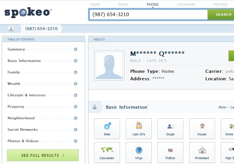
Free Phone Tracer
Provides reverse phone number searches for landlines and cell phones. The owner's name and address is not available for free for cell phones -
only the carrier, line type and phone issuing location is available at
no charge. Owner information for available landline numbers is free.
Users may also track the location of a cell phone number using our reverse directory.
According to the site, it does not use GPS or Wi-Fi tracking to find a cell
phone's current location - search records come from public databases and
directories.

Wireless & Reverse Phone Lookup
Intelius lets you verify phone numbers with this tool. Enter a land-line or mobile phone number to find out if Intelius has information about it. Cell phone numbers return the notation, wireless, while land-based numbers show residential apparently even when they are business numbers.


Phone Lookup Phone Wallpapers Icon Backgrounds Cases Wallpapers Hd Logo Call Numbers Booth Symbol Images Phones

Phone Lookup Phone Wallpapers Icon Backgrounds Cases Wallpapers Hd Logo Call Numbers Booth Symbol Images Phones

Phone Lookup Phone Wallpapers Icon Backgrounds Cases Wallpapers Hd Logo Call Numbers Booth Symbol Images Phones

Phone Lookup Phone Wallpapers Icon Backgrounds Cases Wallpapers Hd Logo Call Numbers Booth Symbol Images Phones
Phone Lookup Phone Wallpapers Icon Backgrounds Cases Wallpapers Hd Logo Call Numbers Booth Symbol Images Phones

Phone Lookup Phone Wallpapers Icon Backgrounds Cases Wallpapers Hd Logo Call Numbers Booth Symbol Images Phones

Phone Lookup Phone Wallpapers Icon Backgrounds Cases Wallpapers Hd Logo Call Numbers Booth Symbol Images Phones

Phone Lookup Phone Wallpapers Icon Backgrounds Cases Wallpapers Hd Logo Call Numbers Booth Symbol Images Phones

Phone Lookup Phone Wallpapers Icon Backgrounds Cases Wallpapers Hd Logo Call Numbers Booth Symbol Images Phones

Phone Lookup Phone Wallpapers Icon Backgrounds Cases Wallpapers Hd Logo Call Numbers Booth Symbol Images Phones

Phone Lookup Phone Wallpapers Icon Backgrounds Cases Wallpapers Hd Logo Call Numbers Booth Symbol Images Phones

Monday, 24 March 2014

Used Cell Phones Phone Wallpapers Icon Backgrounds Cases Wallpapers Hd Logo Call Numbers Booth Symbol Images Phones

Used Cell Phones Biography


Source(google.com.pk)
Researchers are charged up about biobatteries, devices able to harness common biological processes to generate electricity. Most biobatteries are unable to generate large

amounts of power, but researchers recently developed a prototype version that has the potential to be lighter and more powerful than the batteries typically found in today's portable

electronic devices, including smartphones.

In the body, sugar is converted into energy in a process called metabolism, which decomposes sugar into carbon dioxide and water while releasing electrons. Biobatteries produce

energy though the same conversion process by capturing the electrons that are generated in the decomposition of sugar with the same tools that the body uses. Because biobatteries

use materials that are biologically based, they are renewable and non-toxic, making them an attractive alternative to traditional batteries that need metals and chemicals to operate.

Percival Zhang and Zhiguang Zhu, researchers at Virginia Tech, in Blacksburg, designed a new biobattery with a greater output per weight than the typical lithium-ion batteries used in

most electronics. They described the research online last month in the journal Nature Communications.

The new biobattery fully converts sugar to energy, which means more power output than previous biobatteries, and a greater battery charge than common lithium-ion batteries.

“By using the lithium-ion battery, for example, your phone can only last for one day, but in the future it will use sugar as the fuel...then the phone could last 10 days,” said Zhu.

The new biobattery gets its efficiency by using a novel system of enzymes, which are proteins that help the reaction to take place. The system uses two active enzymes that liberate

two pairs of electrons from the sugar, while 10 other enzymes help to reset the reaction inside the biobattery. Once the reaction is reset, the active enzymes release another quartet of

electrons. After six cycles, the biobattery extracts all of the energy bound in the sugar molecule, along with carbon dioxide and water.

Previous biobatteries could only extract one-sixth the energy of the new biobattery, because they didn't use the non-active enzymes for recycling. By extracting more electrons per

weight of sugar, the effective “energy density” of the sugar has increased.

One of the major advantages of this biobattery is that, while the cycle can fully convert sugar to energy, it uses fewer enzymes than the body, making it more robust.

Shelley Minteer, a biobattery expert from the University of Utah in Salt Lake City who was not involved with the work, likes that the team was able to develop an enzyme cycle, also

known as an enzyme pathway, which uses fewer enzymes than the body.

“It’s really important to get all the electrons out, but not just to get all of the electrons out,” said Minteer. She added that it’s important to extract all of the electrons using the fewest

enzymes.

With their new recycling enzyme system, Zhang and his team have done just that. “I think it's a great [enzyme] pathway,” noted Minteer.

While the new enzyme system marks a major step forward for biobatteries, the technology still has some hurdles to surmount before it is market-ready.

“So far there are two more challenges in front of us,” Zhu explained.

He said that, in the current, non-optimized form of the battery, the power output is still too low for many devices and the lifetime of the cell is still too short, as it cannot yet be recharged.

However, as Minteer noted, these challenges are more “on the engineering side of things.” Zhu and Zhang agree and expect to solve these problems at Zhang’s startup company,

Cell-Free Bioinnovations.


Used Cell Phones  Phone Wallpapers Icon Backgrounds Cases Wallpapers Hd Logo Call Numbers Booth Symbol Images Phones

Used Cell Phones  Phone Wallpapers Icon Backgrounds Cases Wallpapers Hd Logo Call Numbers Booth Symbol Images Phones
Used Cell Phones  Phone Wallpapers Icon Backgrounds Cases Wallpapers Hd Logo Call Numbers Booth Symbol Images Phones

Used Cell Phones  Phone Wallpapers Icon Backgrounds Cases Wallpapers Hd Logo Call Numbers Booth Symbol Images Phones

Used Cell Phones  Phone Wallpapers Icon Backgrounds Cases Wallpapers Hd Logo Call Numbers Booth Symbol Images Phones

Used Cell Phones  Phone Wallpapers Icon Backgrounds Cases Wallpapers Hd Logo Call Numbers Booth Symbol Images Phones

Used Cell Phones  Phone Wallpapers Icon Backgrounds Cases Wallpapers Hd Logo Call Numbers Booth Symbol Images Phones

Used Cell Phones  Phone Wallpapers Icon Backgrounds Cases Wallpapers Hd Logo Call Numbers Booth Symbol Images Phones

Used Cell Phones  Phone Wallpapers Icon Backgrounds Cases Wallpapers Hd Logo Call Numbers Booth Symbol Images Phones

Used Cell Phones  Phone Wallpapers Icon Backgrounds Cases Wallpapers Hd Logo Call Numbers Booth Symbol Images Phones

Used Cell Phones  Phone Wallpapers Icon Backgrounds Cases Wallpapers Hd Logo Call Numbers Booth Symbol Images Phones

411 Reverse Phone Phone Wallpapers Icon Backgrounds Cases Wallpapers Hd Logo Call Numbers Booth Symbol Images Phones

411 Reverse Phone Biography


Source(google.com.pk)

Background

Facioscapulohumeral muscular dystrophy (FSHD) is an autosomal dominant muscle disorder, which is linked to the contraction of the D4Z4 array at chromosome 4q35. Recent

studies suggest that this shortening of the D4Z4 array leads to aberrant expression of double homeobox protein 4 (DUX4) and causes FSHD. In addition, misregulation of microRNAs

(miRNAs) has been reported in muscular dystrophies including FSHD. In this study, we identified a miRNA that is differentially expressed in FSHD myoblasts and investigated its

function.

Methods

To identify misregulated miRNAs and their potential targets in FSHD myoblasts, we performed expression profiling of both miRNA and mRNA using TaqMan Human MicroRNA Arrays

and Affymetrix Human Genome U133A plus 2.0 microarrays, respectively. In addition, we over-expressed miR-411 in C2C12 cells to determine the effect of miR-411 on myogenic

markers.

Results

Using miRNA and mRNA expression profiling, we identified 8 miRNAs and 1,502 transcripts that were differentially expressed in FSHD myoblasts during cell proliferation. One of the

8 differentially expressed miRNAs, miR-411, was validated by quantitative RT-PCR in both primary (2.1 fold, p<0.01) and immortalized (2.7 fold, p<0.01) myoblasts. In situ

hybridization showed cytoplasmic localization of miR-411 in FSHD myoblasts. By analyzing both miRNA and mRNA data using Partek Genomics Suite, we identified 4 mRNAs

potentially regulated by miR-411 including YY1 associated factor 2 (YAF2). The down-regulation of YAF2 in immortalized myoblasts was validated by immunoblotting (−3.7 fold,

p<0.01). C2C12 cells were transfected with miR-411 to determine whether miR-411 affects YAF2 expression in myoblasts. The results showed that over-expression of miR-411

reduced YAF2 mRNA expression. In addition, expression of myogenic markers including Myod, myogenin, and myosin heavy chain 1 (Myh1) were suppressed by miR-411.

Conclusions

The study demonstrated that miR-411 was differentially expressed in FSHD myoblasts and may play a role in regulating myogenesis.

Keywords: FSHD, microRNA, miR-411, YAF2, YY1, Myod, myogenin
Go to:
Background
Facioscapulohumeral muscular dystrophy (FSHD) is an autosomal dominant myopathy with estimated prevalence of 1:20,000 [1,2]. The age of onset is often in the second decade of

life with nearly complete penetrance (95%) by age 20 [3]. FSHD is characterized by progressive weakness of different muscle groups, often starting with the facial muscles, followed

by the shoulder girdle muscles, and moving down to the hip girdle and the extremities [4-6]. Many patients also exhibit a marked left-right asymmetry in muscle involvement [4,7,8].

Patients can have additional symptoms such as severe inflammation in muscles, subclinical hearing loss, and peripheral retinal capillary abnormalities [9-11]. Genetic studies of

FSHD have shown that the disease is associated with a deletion of the D4Z4 repeats in the 4q35 subtelomeric region. In individuals without FSHD, this region contains up to 150

copies of the D4Z4 repeats while patients with FSHD only have one to ten copies of the repeat [4,6,12-16].

Each D4Z4 repeat contains a double homeobox protein 4 (DUX4) gene. Several DUX4 splice variants have been reported to be expressed in germ-line cells and myoblasts [17-19].

Although the function of DUX4 is not yet known, the full-length DUX4 transcript (fl-DUX4) has been shown to be cytotoxic in vivo and ex vivo when ectopically expressed [19-23].

Several studies suggested that p53-dependent cell death plays a major role in the cytotoxicity of DUX4 [23-25]. Recent studies have also shown that a combination of two genomic

features is required to cause FSHD. First, the contraction of the D4Z4 repeats causes hypomethylation of the D4Z4 region, allowing DUX4 mRNA to be transcribed [21,26]. Second,

an intact polyadenylation signal in the region distal to the D4Z4 array allows DUX4 transcripts from the last D4Z4 repeat to be polyadenylated and therefore stable for protein

translation. This combination of events leads to the aberrant expression of DUX4 and the downstream molecular changes involved in FSHD [17,21,27]. The aberrant expression of

DUX4 in FSHD has been proposed to inhibit myogenesis by suppressing Myod regulated pathways and inducing muscle atrophy pathways [20,24,28-33]. However, the regulatory

relationship between DUX4 and these pathways is not clear. Currently no effective therapy for FSHD is available. Several pharmacological treatments such as corticosteroids,

albuterol, creatine monohydrate, and anti-human myostatin antibody have been tested for their efficacy of treating FSHD, but none showed promising results [34].

MicroRNAs (miRNAs) are short (~22 nucleotides) non-coding RNAs which regulate gene expression by interfering translation or promoting degradation of target mRNAs [35,36]. A

mature miRNA is generated through several steps. First, a primary-miRNA (pri-RNA) is transcribed and then cleaved to form a pre-miRNA, which is a single hairpin-shaped stem-loop

[37]. Subsequently, the pre-miRNA is exported to the cytoplasm and cleaved into a mature miRNA duplex by Dicer [37,38]. The functional strand is incorporated into the RNA-induced

silencing complex (RISC) to form a miRNA-RISC complex [39,40]. In general, the miRNA-RISC complex will cleave the target mRNA when the target sequence is perfectly

complementary to the miRNA sequence, or it will interfere with translation of the target mRNA when mismatches are present in the target sequence [40,41]. Many miRNAs are

conserved between vertebrates and invertebrates and have been shown to share functions in various cellular processes including embryogenesis, organogenesis, apoptosis, cell

cycle regulation and disease development, including muscle disorders [40-43]. Several miRNAs have been shown to play important roles in muscle differentiation, including the miR-1

and miR-133 families, miR-181, miR-214, miR-24, miR-221 and miR-222 [44-51]. In additional to normal muscle growth and maintenance, miRNAs have also been shown to be

differentially expressed in disease conditions [52-54]. A global miRNA expression profile of 10 muscle disorders was previously performed and showed that 185 out of the 428

miRNAs examined were differentially expressed in at least one of the 10 different muscle disorders [55]. Among the 185 miRNAs, 62 were up-regulated in FSHD while none was

down-regulated. These findings suggest that miRNAs may play a critical role in FSHD, although the mechanisms involved have not been studied. MiR-411 belongs to the miR-379

family and is located in the miR-379/miR-656 cluster within the DLK-DIO3 region on human chromosome 14 [56]. The miR-379/miR-656 cluster is highly conserved in placental

mammals [56]. In mouse brain, the expression of the miR-379/miR-656 gene cluster is likely co-regulated by myocyte enhancing factor 2 (Mef2) and is involved in activity-dependent

outgrowth of hyppocampal neurons [57]. The function of miR-411 in brain or other tissues is currently unknown. In this study we performed miRNA expression profiling using PCR-

based miRNA arrays to identify miRNAs misregulated in FSHD myoblasts. We then used mRNA profiling to identify potential regulatory targets of miR-411, which was significantly

upregulated in the miRNA profiling. We further examined the effects of over-expressing miR-411 in C2C12 myoblasts and its potential role in myogenesis.

Go to:
Methods
Cell culture and immunostaining

Primary myoblasts were obtained from EuroBioBank (Dr. Schneiderat and Dr. Walter) (Additional file 1: Table S1). For expression profiling experiments, cells were cultured in

collagen I-coated flasks with SkGM (Lonza) at 37°C, 5% CO2. For in situ hybridization experiments, cells were seeded on poly-D-lysine/mouse laminin-coated coverslips (BD

BioCoat, BD Biosciences).

C2C12 cells were purchased from ATCC and cultured in growth medium consisting of DMEM (Life Technologies) with 10% heat-inactivated fetal bovine serum (Sigma). Myotube

differentiation was induced by culturing the cells in differentiation medium consisting of DMEM with 2% heat-inactivated horse serum (Sigma) at 37°C, 5% CO2.

Immortalized human myoblasts were from the Boston Biomedical Research Institute and cultured as described in previously published protocol [58,59]. Briefly, immortalized myoblasts

were cultured in a growth medium consisting of medium 199 and DMEM (Life Technologies) in a 1:4 ratio with 0.8 mM sodium pyruvate (Life Technologies), 3.4 g/l sodium

bicarbonate (Sigma-Aldrich), 15% fetal bovine serum (Thermo Scientific), 0.03 μg/ml Zinc sulfate (Fisher), 1.4 μg/ml vitamin B12 (Sigma-Aldrich), 2.5 ng/ml recombinant human

hepatocyte growth factor (Millipore), 10 ng/ml basic fibroblast growth factor (BioPioneer), 0.02 M HEPES (Life Technologies), and 0.055 μg/ml dexamethasone (Sigma-Aldrich) at

37°C, 5% CO2. The culture dish was coated with 0.1% gelatin (Sigma-Aldrich).

Myoblasts purity was determined by performing immunofluorescent staining using anti-human desmin (Dako) antibody. Myoblasts that exhibited greater than 70% desmin-positive

cells were utilized for expression profiling. Immunostaining was conducted as previously described [60]. Briefly, the cells were fixed with 4% paraformaldehyde for 30 minutes, then

blocked with 0.3% Triton X-100, 15% hose serum and 450 mM NaCl in phosphate buffer saline (PBS). Following blocking, fixed cells were incubated with anti-human desmin (Dako)

for 4°C overnight. After washing 3 times, fixed cells were incubated with secondary antibody, DyLight 488-conjugated donkey anti-mouse IgG (Jackson ImmunoRessearch

Laboratories). The slides were mounted using ProLong Gold Antifade Reagent with DAPI (Life Technologies) for further examination.

Total RNA extraction and miRNA expression profiling

To identify differentially expressed miRNA in FSHD primary myoblasts, we performed miRNA expression profiling using proliferating primary FSHD and control myoblasts (n=3,

Additional file 1: Table S1). Total RNA with miRNA enrichment was extracted from cells using mirVana miRNA isolation kit (Life Technologies) according to manufacturer’s protocol.

Following RNA isolation, RNA quality and concentration were determined by gel electrophoresis and NanoDrop (Thermo Fisher Scientific), respectively. The miRNA profile of each

myoblasts was determined using TaqMan Human MicroRNA Array v2.0 (Human Array A) (Life Technologies) according to manufacturer’s protocol. Briefly, reverse transcription (RT)

was performed with 100 ng of total RNA, Multiplex RT Human primer pools, and TaqMan MicroRNA Reverse Transcriptase Kit (Life Technologies). Real-time PCR was performed with

TaqMan Universal PCR Master Mix, No AmpErase UNG (Life Technologies) using the Applied Biosystems 7900HT System. Ct values of all miRNAs were determined using RQ

Manager 1.2 (Life Technologies) with a threshold of 0.1. Ct values were then imported into Partek Genomics Suite 6.5 and normalized to control, RNU48. ANOVA analysis was

performed using Partek Genomics Suite 6.5 (Partek Incorporated, MO). No multiple testing corrections were performed.


411 Reverse Phone  Phone Wallpapers Icon Backgrounds Cases Wallpapers Hd Logo Call Numbers Booth Symbol Images Phones

411 Reverse Phone  Phone Wallpapers Icon Backgrounds Cases Wallpapers Hd Logo Call Numbers Booth Symbol Images Phones

411 Reverse Phone  Phone Wallpapers Icon Backgrounds Cases Wallpapers Hd Logo Call Numbers Booth Symbol Images Phones

411 Reverse Phone  Phone Wallpapers Icon Backgrounds Cases Wallpapers Hd Logo Call Numbers Booth Symbol Images Phones

411 Reverse Phone  Phone Wallpapers Icon Backgrounds Cases Wallpapers Hd Logo Call Numbers Booth Symbol Images Phones

411 Reverse Phone  Phone Wallpapers Icon Backgrounds Cases Wallpapers Hd Logo Call Numbers Booth Symbol Images Phones

411 Reverse Phone  Phone Wallpapers Icon Backgrounds Cases Wallpapers Hd Logo Call Numbers Booth Symbol Images Phones

411 Reverse Phone  Phone Wallpapers Icon Backgrounds Cases Wallpapers Hd Logo Call Numbers Booth Symbol Images Phones

411 Reverse Phone  Phone Wallpapers Icon Backgrounds Cases Wallpapers Hd Logo Call Numbers Booth Symbol Images Phones

411 Reverse Phone  Phone Wallpapers Icon Backgrounds Cases Wallpapers Hd Logo Call Numbers Booth Symbol Images Phones
411 Reverse Phone  Phone Wallpapers Icon Backgrounds Cases Wallpapers Hd Logo Call Numbers Booth Symbol Images Phones

All Phones Phone Wallpapers Icon Backgrounds Cases Wallpapers Hd Logo Call Numbers Booth Symbol Images Phones
All Phones Biography


Source(google.com.pk)
BLACK Cell Phone Radiation Shield - Anti-Radiation Protector Sticker for... Bio Shield - Protector for EMF and Wi-Fi RADIATION, creates energy VORTEX WHICH RESONATES

AND BOOSTS BODY ENERGY, Provides Natural Immunity to Wi-Fi, GUARANTEED PROTECTION, SEE TESTIMONIALS ON YOUTUBE FOR vORTEXbIOsHIELD, immediately

Effective - Right away NO HEATING ON THE CHEEK. OFFERS A COMPLETELY POSITIVE AND HEALTHY EFFECT ON YOU. WORKS WITH ALL PHONES, COMPATIBLE WITH

IPHONE OR ANY OTHER PHONE - PERSONAL RADIATION PROTECTOR VORTEX BIO SHIELD ATTACHES TO THE PHONE OR PHONE CASE (RECOMMENDED). NO

DAMAGE TO THE SURFACE, ATTACHES IN SECONDS WITH DOUBLE SIDED HEAVY DUTY TAPE TO THE PHONE CASE

Mobile Phones
'Casualty catastrophe': cell phones and child brains
| Print |  Email
RT Television, 20 October 2013

Insurers stop covering for cell phone use, called the next 'casualty catastrophe' after tobacco and asbestos; phone manufacturers hit with a class action and personal lawsuits; and the

warning deep inside your mobile.

Seek truth from facts with Ellie Marks, whose husband Alan is suing the industry for his brain tumor, 'cell phone survivor' Bret Bocook, leading radiation biologist Prof. Dariusz

Leszczynski, Microwave News editor Dr. Louis Slesin, Storyleak editor Anthony Gucciardi, and former senior White House adviser Dr. Devra Davis.

LTE Cell Phone Radiation Affects Brain Activity in Cell Phone Users
| Print |  Email
PRLog (Press Release), 23 September 2013

The first study on the short-term effects of Long Term Evolution (LTE), the fourth generation cell phone technology, has been published online in the peer-reviewed journal, Clinical

Neurophysiology. (1)

In a controlled experiment, researchers exposed the right ear of 18 participants to LTE cellphone radiation for 30 minutes. The source of the radiation was 1 centimeter from the ear,

and the absorbed amount of radiation in the brain was well within international (ICNIRP) cell phone legal limits. The researchers employed a double-blind, crossover, randomized and

counter-balanced design to eliminate any possible study biases.

The resting state brain activity of each participant was measured by magnetic resonance imaging (fMRI) at two times -- after exposure to LTE microwave radiation, and after a sham

exposure.

The results demonstrated that LTE exposure affected brain neural activity not only in the closer brain region but also in the remote region, including the left hemisphere of the brain. The

study helps explain the underlying neural mechanism for the remote effects of microwave radiation in the brain.


All Phones  Phone Wallpapers Icon Backgrounds Cases Wallpapers Hd Logo Call Numbers Booth Symbol Images Phones

All Phones  Phone Wallpapers Icon Backgrounds Cases Wallpapers Hd Logo Call Numbers Booth Symbol Images Phones

All Phones  Phone Wallpapers Icon Backgrounds Cases Wallpapers Hd Logo Call Numbers Booth Symbol Images Phones

All Phones  Phone Wallpapers Icon Backgrounds Cases Wallpapers Hd Logo Call Numbers Booth Symbol Images Phones

All Phones  Phone Wallpapers Icon Backgrounds Cases Wallpapers Hd Logo Call Numbers Booth Symbol Images Phones

All Phones  Phone Wallpapers Icon Backgrounds Cases Wallpapers Hd Logo Call Numbers Booth Symbol Images Phones

All Phones  Phone Wallpapers Icon Backgrounds Cases Wallpapers Hd Logo Call Numbers Booth Symbol Images Phones

All Phones  Phone Wallpapers Icon Backgrounds Cases Wallpapers Hd Logo Call Numbers Booth Symbol Images Phones

All Phones  Phone Wallpapers Icon Backgrounds Cases Wallpapers Hd Logo Call Numbers Booth Symbol Images Phones

All Phones  Phone Wallpapers Icon Backgrounds Cases Wallpapers Hd Logo Call Numbers Booth Symbol Images Phones

All Phones  Phone Wallpapers Icon Backgrounds Cases Wallpapers Hd Logo Call Numbers Booth Symbol Images Phones